
Overall goal and technology
In the Pfisterer Lab we investigate the regulatory mechanisms that maintain tissue homeostasis and how their dysregulation drives inflammatory disease and cancer. Our research sits at the interface of immunology, cell biology, and tissue mechanics.
We combine advanced microscopy, next-generation sequencing, and molecular, biochemical and cellular biology approaches to study tissue regulation across scales. By integrating these technologies, we aim to understand how cellular states, signaling networks, and the physical properties of tissues interact to maintain homeostasis or drive pathology.
PhotoMel
Malignant melanoma is a leading cause of cancer mortality among young Europeans. This persistence indicates that factors beyond UVB-induced DNA damage contribute to risk. In this project we aim to investigate how early-life UVA and chemical exposure remodel the skin microenvironment to drive melanoma initiation and early spread.
In this multi-consortium EU project, coordinated by Karin Pfisterer, we will integrate ex vivo skin models, mutational characterisation, extracellular matrix biomechanics, spatial multiomics, advanced microscopy, CROP-seq functional genomics and multiscale computational modelling in collaboration with CeMM, King’s College London, Leiden University, EMBL Heidelberg, Marije Kruis, Region Skäne and Lund University.
Our objectives are to
- prevent melanoma onset by decoding how chemical exposures modulate UVA-induced skin damage
- map patient biopsy data to resolve spatial tumour-microenvironment interactions
- transform mechanistic insights into predictive 3D computational models;
- predict patient-specific metastatic risk through biomarker discovery; and
- engage citizens and patients to restore trust, guide regulation and strengthen prevention strategies.
Specifically, we will quantify UVA-chemical effects on skin cells and the ECM, define how altered microenvironments shape melanoma subpopulations, and integrate ECM, immune and tumour features into predictive models, delivering biomarker panels for exposure-driven metastatic risk stratification in young adults under 40 years.
Pioneer melanoma cells
Cancer metastasis is the reason why later stages of melanoma are closely associated with poor patient prognosis. To metastasize, cells migrate from the initial tumor into the surrounding tissue and even distal sites in the body. Cells sense and interact with the extracellular matrix, mainly through cell protrusions called filopodia. To migrate, epithelial melanoma cells undergo various changes to transdifferentiate into a mesenchymal phenotype, enabling migration and metastasis. From a primary tumor, few initial highly migratory cells, pioneer melanoma cells, are hypothesized to initiate invasion of the surrounding tissue. The migration tracks that form in the fibers of the extracellular matrix enable cells to follow the initial pioneer cancer cells, thereby facilitating metastasis formation.
The project aims to identify the changes cells undergo when switching from an epithelial to mesenchymal phenotype. In this project, we focus on detecting the interaction of cancer cells with their environment which plays a crucial role in invasiveness.
Collagen matrices of varying stiffness are used to mimic the native environment of melanoma cells in the skin. Through live cell imaging of these cells in 3D matrices, we may better understand the mechanisms behind their invasive behavior. Further, gene expression will be analyzed to investigate the epithelial-to-mesenchymal transition at the molecular level.
People involved in the projects: Karin Pfisterer (Principal Investigator), Pauline Weinzettl
ECM modifications in inflammatory skin diseases
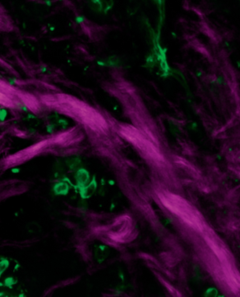
A major focus of the Pfisterer Lab is understanding how extracellular matrix (ECM) remodeling and tissue mechanics shape inflammatory tissue states in human skin. The skin ECM is not merely a structural scaffold but a dynamic signaling environment that regulates immune cell behavior, tissue integrity, and cellular communication. During chronic inflammation, these regulatory networks become profoundly altered, leading to changes in matrix architecture, mechanical properties, and cellular function.
Using advanced imaging, single-cell transcriptomics, and mechanobiology approaches, we investigate how fibroblasts and immune cells cooperate to remodel the ECM in inflammatory skin diseases such as psoriasis. Our recent work identified striking alterations in collagen organization within psoriatic skin, including aligned and stiffened collagen bundles associated with elevated expression of the matrix-crosslinking enzyme lysyl oxidase (LOX). We further uncovered a mechanosensitive HIF-1-LOX regulatory axis in dermal fibroblasts, linking tissue stiffness to ECM remodeling and inflammatory tissue adaptation.
In our ongoing research we aim to identify the mechanisms by which altered tissue mechanics actively contribute to disease progression by reinforcing chronic inflammatory states through mechano-inflammatory feedback loops. By defining how mechanical signals, ECM composition, and cellular states interact, we aim to uncover novel therapeutic vulnerabilities in chronic inflammatory disease.
People involved in the projects: Karin Pfisterer (Principal Investigator), Parvaneh Balsini, David Samardzic, Pauline Weinzettl